
Health
TB deaths decline in India, but diagnostic gaps threaten elimination goals
India has reduced tuberculosis mortality by 38% since 2015, according to the WHO Global Tuberculosis Report 2026. However, gaps in diagnostic infrastructure, particularly in drug-resistance testing and laboratory facilities, continue to challenge the country’s TB elimination efforts.


India has recorded a 38% decline in tuberculosis (TB) mortality since 2015, outperforming the global reduction of 35%. However, significant gaps in diagnostic infrastructure, particularly at primary healthcare facilities, could complicate the country’s efforts to eliminate the disease.
The findings present a mixed picture of India’s TB response. The World Health Organization’s (WHO) Global Tuberculosis Report 2026, released on November 12, documents improvements in mortality and treatment outcomes. At the same time, a multi-state study published in the journal Microbiology Spectrum in August 2026 identifies substantial shortages in essential diagnostic services, including facilities required to detect drug-resistant TB.
According to the WHO report, India’s estimated TB mortality rate declined from 28 deaths per lakh population in 2015 to 18 per lakh in 2025. The country also recorded a 94% treatment success rate among patients with drug-susceptible TB.
The figures indicate progress in early detection, treatment access and patient support. However, reducing mortality does not necessarily mean that diagnostic services are equally accessible across the country’s healthcare system.
Diagnostic infrastructure remains inadequate
The study, titled Evaluating the nationwide availability of essential diagnostics for tuberculosis toward achieving a TB-free India, examined 327 public healthcare facilities across seven states and Delhi. It assessed the availability of diagnostic services recommended under India’s National Essential Diagnostics List.
One of its major findings was a 61.56% shortfall in culture and drug-susceptibility testing (CDST) laboratory capacity compared with population-based requirements. The researchers identified only 97 CDST laboratories against 6,308 TB Treatment Units.
Drug-susceptibility testing is essential for identifying whether tuberculosis bacteria are resistant to particular medicines. Without timely testing, patients with drug-resistant TB may face delays in receiving appropriate treatment, potentially increasing the risk of continued transmission and unsuccessful treatment outcomes.
The study also found that only 4.94% of the surveyed Primary Health Centres (PHCs) had the complete set of essential TB diagnostic facilities recommended for that level of healthcare.
Across the assessed facilities, cartridge-based nucleic acid amplification testing (CBNAAT), which can rapidly detect TB and certain forms of drug resistance, was available in 51.28%. X-ray services were also available in 51.28% of the facilities.
The researchers emphasised that expanding access to individual diagnostic technologies would not be sufficient without trained personnel, functional laboratory networks, maintenance, quality assurance and effective referral systems.
The findings should be interpreted as a multi-state assessment rather than a nationally representative survey of every public healthcare facility. Nevertheless, they identify infrastructure weaknesses that require further attention.
Screening expands across the country
India has expanded its screening initiatives under the TB Mukt Bharat Abhiyaan, which focuses on identifying undiagnosed cases, particularly among vulnerable populations.
According to the Union Ministry of Health and Family Welfare, more than 25 crore people were brought under intensified screening activities between December 2024 and November 8, 2026. The programme conducted over 11 crore chest X-rays and more than three crore molecular diagnostic tests.

A healthcare professional examining a chest X-ray.Image credit:Pexel
The campaign identified 48.64 lakh new TB cases, including 14.97 lakh asymptomatic cases. Detecting individuals without noticeable symptoms is particularly important because undiagnosed infections can delay treatment and contribute to transmission.
The government has also expanded nutritional assistance through the Ni-kshay Poshan Yojana and community support through the Ni-kshay Mitra initiative. These interventions recognise that TB treatment involves more than medication, particularly for patients experiencing undernutrition and financial difficulties.
Drug resistance and the challenge ahead
Drug-resistant tuberculosis remains an important concern for public health systems. Treating drug-resistant infections requires accurate diagnosis and appropriate treatment regimens, making access to comprehensive laboratory services essential.
The diagnostic study argues that rapid molecular tests must be complemented by functional culture-based testing facilities capable of identifying a broader range of drug resistance. It also highlights the potential of next-generation sequencing for resistance profiling, although costs, infrastructure and bioinformatics capacity remain barriers to wider implementation.
The WHO report, which draws on information from 187 countries and territories covering more than 99% of the global population and TB cases, provides an important assessment of progress towards international TB targets.
India’s declining mortality and improved treatment outcomes represent significant gains. However, sustaining this progress will depend on whether diagnostic services can reach patients before the disease advances and whether healthcare facilities can identify drug resistance promptly.
As India continues its TB elimination efforts, strengthening primary healthcare laboratories, expanding molecular testing and improving access to drug-susceptibility testing will be critical. The next phase of progress must focus not only on screening more people but also on ensuring that every suspected case can access accurate diagnosis and appropriate treatment.



Space & Physics
Can Studying Cancer Tumours in Space Help Improve Chemotherapy on Earth?
Researchers are studying miniature models of colorectal cancer aboard the International Space Station to understand whether microgravity changes how cancer cells respond to chemotherapy.

Two people diagnosed with the same type of cancer may not respond to the same treatment in the same way. A chemotherapy drug that works well for one patient may be less effective for another. This happens partly because tumours differ in their genetic makeup, growth patterns and response to medicines. Understanding these differences is vital for developing cancer research and treatments that work better for individual patients.
By sending miniature models of cancer tumours into space, scientists at Wake Forest Institute for Regenerative Medicines in the United States are exploring an unusual way to explore the problem. They are investigating whether the absence of normal gravity changes how colorectal cancer cells respond to chemotherapy. The project, called Tumor Avatars in Space (TAVIS), uses the International Space Station (ISS) as a laboratory to study how cancer cells behave in microgravity.
The experiment uses organoids, which are small, three-dimensional models of human tissues grown in laboratories. For this project, researchers developed tumour organoids using cancer cells collected from patients with colorectal cancer. These miniature models retain some of the biological features of the original tumours, allowing scientists to study how they respond to medicines.
The researchers will compare how these tumour models respond to chemotherapy in space and on Earth. By studying the differences, they hope to understand whether microgravity changes the way cancer cells react to treatment.
Why Study Cancer Cells in Space?
Gravity influences how cells grow and arrange themselves. On Earth, cells are affected by gravitational forces and the way they settle within their surroundings. In microgravity, these conditions change, allowing cells to form three-dimensional structures in different ways. This makes space an interesting environment for studying how cells behave. Researchers want to know whether changes in these conditions can affect tumour growth, cell activity and responses to cancer drugs.
Cancer tumours are made up of more than just cancer cells. They contain different types of cells that interact with one another and their surroundings. These interactions can influence how a tumour grows and responds to treatment. Studying three-dimensional tumour models can help scientists understand some of these processes better.
In the TAVIS experiment, researchers will examine whether colorectal cancer cells become more sensitive to chemotherapy under microgravity conditions. If they find differences, they will investigate the genetic and cellular changes responsible for them. One area of interest is how cancer cells use energy. This process, known as metabolism, helps cells grow, survive and carry out their functions. Changes in metabolism could influence how effectively chemotherapy destroys cancer cells.
The researchers hope to identify the biological processes behind any changes in treatment response. They will then investigate whether similar effects can be reproduced on Earth, potentially by targeting particular genes or cellular processes.
What are Tumour Organoids?
Organoids are miniature, three-dimensional structures grown in laboratories using stem cells or cells taken from human tissues. Depending on the cells used, scientists can grow models that reproduce certain features of organs such as the intestine, liver, brain and lungs. In cancer research, scientists can use cells collected from a patient’s tumour to grow patient-derived tumour organoids. These models retain some of the characteristics of the original cancer and allow researchers to test how different medicines affect it.
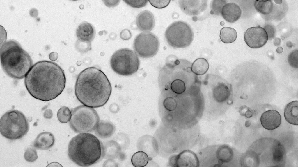
This approach provides more information than experiments using cancer cells grown in a flat layer in laboratory dishes. Since organoids have a three-dimensional structure, they can better represent some of the conditions in which cells grow and interact inside the body.
Researchers are also studying whether patient-derived organoids could help doctors identify treatments that are more likely to work for individual patients. However, this approach still requires further research and is not yet a replacement for established clinical treatment decisions.
Can it Help Cancer Treatment?
The TAVIS project is part of a wider effort to use space-based research to understand human biology and develop medical applications on Earth. After the experiment, researchers will compare the space-grown tumour models with those maintained on Earth. They will examine whether microgravity caused changes in the cancer cells and whether those changes affected their response to chemotherapy.
“If microgravity makes these tumors more responsive, we can look for the genetic changes behind that and try to induce the same changes here on Earth,” said Shay Soker, Ph.D., WFIRM professor and principal investigator of the study. “It’s similar in spirit to immunotherapy, which makes tumors more visible to the immune system. The tumor can run, but it can’t hide.”
If the space-grown models respond differently, scientists will investigate whether the same effects can be produced under normal conditions on Earth. This could help identify new ways to make cancer cells more responsive to existing treatments.
The experiment does not mean that cancer can be treated more effectively in space. Instead, it uses the unusual conditions aboard the ISS to investigate biological changes that are difficult to study in conventional laboratories. By understanding how cancer cells respond to different conditions, researchers hope to gain new insights into tumour behaviour, drug development and the possibility of more personalised cancer treatment.
Health
Over 1,000 Cancer Drug Formulations Become Cheaper Across India
India has reduced prices across 1,031 anti-cancer medicine formulations, with the government estimating annual consumer savings of 3,502 crore rupees.
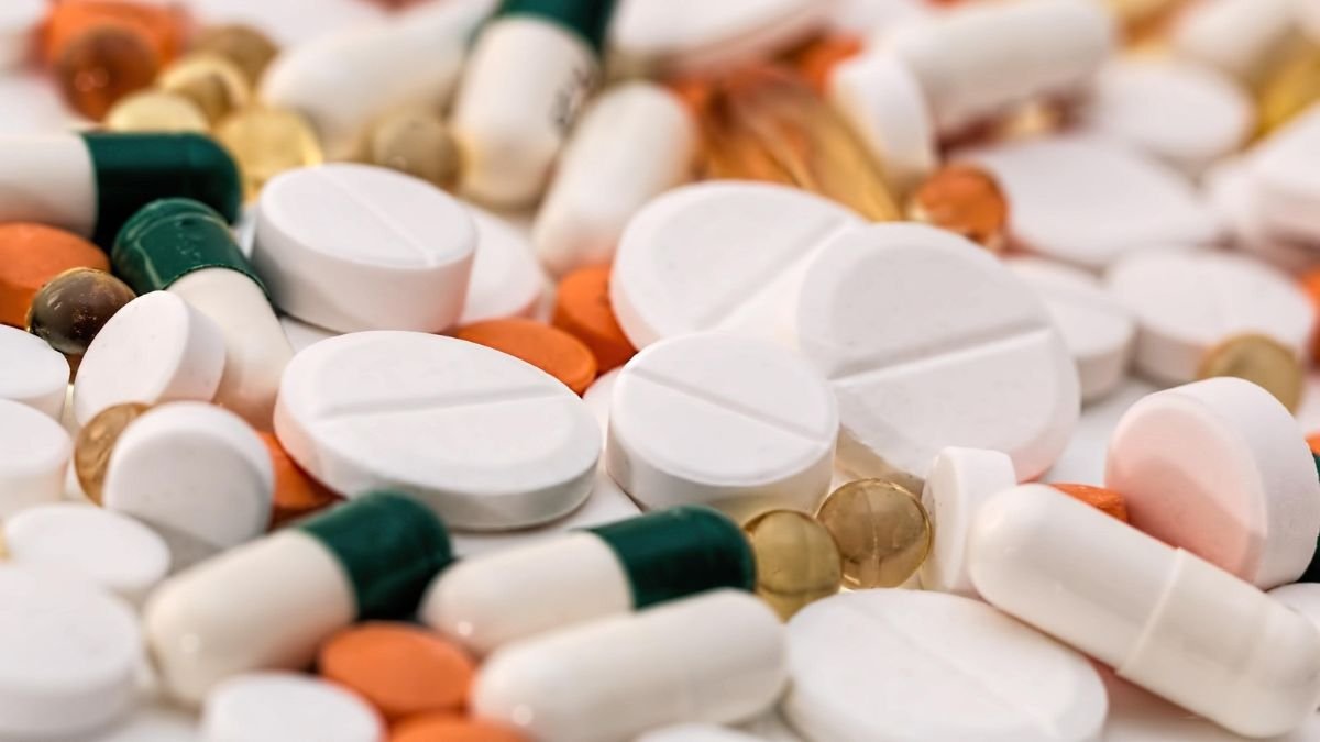
Cancer treatment can involve months or even years of medication, repeated diagnostic tests, hospital visits and specialised procedures. For many Indian families, the financial strain continues long after the initial diagnosis. Against this backdrop, a government intervention to reduce the prices of more than 1,000 anti-cancer drug formulations has come into effect on November 9. The National Pharmaceutical Pricing Authority (NPPA), under the Department of Pharmaceuticals, has rationalised trade margins on 137 identified non-scheduled anti-cancer drugs. The government estimates that the revised prices will save consumers 3,502.31 crore rupees annually.
According to the Ministry of Chemicals and Fertilizers, 76 pharmaceutical companies have reported revised maximum retail prices (MRPs) for 1,225 formulations covering 121 drugs. Of these, 1,031 formulations have registered price reductions. Around 600 brands have reported reductions exceeding 50%, while the highest reduction stands at 96.45%.
The intervention is significant given the scale of cancer in India. The Indian Council of Medical Research’s National Cancer Registry Programme estimated 15.69 lakh new cancer cases in 2025. Breast, lung and mouth cancers featured among the leading cancer sites.
What has Changed in Medicine Pricing?
The latest intervention focuses on non-scheduled medicines, which are not subject to the same ceiling-price mechanism as drugs included in the National List of Essential Medicines.
The Drugs (Prices Control) Order, 2013, gives the government the power to control medicine prices when necessary to protect public interest. It fixes maximum prices for scheduled medicines, while prices of non-scheduled medicines are generally monitored. Using this order, the government has now stepped in to limit the trade margins on the selected anti-cancer medicines.
Trade margins represent the difference between the price at which medicines are supplied through the distribution chain and the price at which they are sold to consumers. Excessive mark-ups can make medicines significantly more expensive before they reach patients. Under the new arrangement, the trade margin on the identified medicines has been capped at 30% of the MRP. The measure follows a similar exercise in 2019, when trade margins on 42 non-scheduled anti-cancer medicines were rationalised.
The revised prices cover medicines used in treating different cancers. These include Bevacizumab, used for lung, colon, ovarian and cervical cancers; Denosumab, used in breast and prostate cancer patients to prevent bone loss; and Pegfilgrastim, prescribed to prevent neutropenia in cancer patients.
The price changes are substantial for several brands. Zydus Lifesciences’ Bevacizumab brand Bryxta, for instance, has seen its MRP fall from 25,995 rupees to 3,318 rupees. Reliance Life Sciences’ Abiraterone acetate brand Abirone has declined from 41,250 rupees to 3,218 rupees. Similarly, the price of Reliance Life Sciences’ Peg-ReliGrast has fallen from 5,331 rupees to 1,258 rupees.
Cancer Drugs: Financial Burden of Cancer Care
The importance of reducing medicine prices becomes clearer when viewed against the financial burden of cancer treatment. A study published in Frontiers in Public Health in 2023, involving 12,148 cancer patients across seven treatment centres in India, estimated the average annual direct out-of-pocket expenditure on cancer treatment at 3.31 lakh rupees per patient. Medicines accounted for 45% of expenditure during hospitalisation, while diagnostics contributed 36.4% of outpatient treatment expenditure.

The findings illustrate why medicine affordability matters, particularly for patients who require repeated treatment. Even when the cost of a particular drug declines, expenses related to diagnostics, surgery, hospitalisation and travel can continue to weigh heavily on households.
What Happens Next?
The revised prices will apply to the identified medicines, including 10–15 drugs that have not yet been launched in India. Their prices will also be subject to the margin restrictions when they enter the market. Manufacturers can undertake annual price increases in accordance with the Drugs (Prices Control) Order, 2013, subject to continued compliance with the maximum permissible trade margin.
The NPPA has also directed retailers, dealers, hospitals and medical institutions to prominently display the revised MRP lists. State Drug Controllers will monitor compliance. Consumers can verify medicine prices through the Pharma Sahi Daam mobile application and the NPPA website. The reduction addresses an important component of cancer treatment expenditure: the price patients pay for medicines. Its impact, however, will depend on whether the revised prices are implemented consistently across pharmacies and hospitals, and whether patients can access the medicines they need.
Lower medicine prices can ease a substantial part of the financial burden. Making cancer treatment affordable, however, will also require attention to diagnostic costs, hospital charges and access to quality treatment across the public healthcare system.
Health
How Rubella Virus Finds Its Way Into the Placenta: Scientists Identify a Human Cell Receptor
A new Nature study identifies Nectin-4 as a receptor that helps the rubella virus enter placental and respiratory cells, offering insight into congenital rubella syndrome.
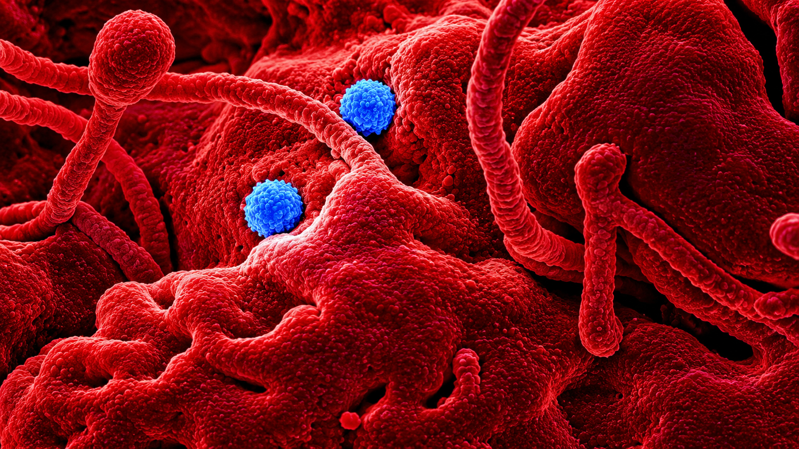
An infection that causes little more than a rash and mild fever in many people can have devastating consequences when it occurs during pregnancy. Rubella, a contagious viral disease, can cross the maternal–foetal interface and interfere with the development of an unborn child, causing hearing loss, heart defects, cataracts and other complications collectively known as congenital rubella syndrome (CRS).
An estimated 26,859 children were born with congenital rubella syndrome worldwide in 2024, according to a modelling analysis published by the US Centers for Disease Control and Prevention (CDC) in July 2026. Although this represents a substantial decline from an estimated 101,381 cases in 2012, the disease continues to affect newborns in countries where vaccination coverage remains inadequate. But how does the rubella virus enter the cells lining the respiratory tract and the placenta?
A study published in Nature identifies a protein called Nectin-4 as one of the cellular entry points used by the virus. The finding offers a closer look at how rubella establishes infection in two tissues central to its transmission and its effects during pregnancy.

Protein That Opens the Door
Like other viruses, rubella cannot reproduce on its own. It must enter a living cell and use its machinery to produce new viral particles. This often begins when proteins on the virus’s outer envelope bind to specific molecules on the surface of a human cell. These molecules, called receptors, determine which cells a virus can enter. Identifying them helps researchers understand why certain tissues are vulnerable to infection.
The research team found that rubella uses Nectin-4, a cell-adhesion protein, to gain entry into respiratory and placental cells. Nectin-4 normally helps neighbouring cells maintain contact with one another and is present in several human tissues. The virus carries two envelope proteins, E1 and E2, which participate in its interaction with host cells. The researchers found that this viral protein complex interacts directly with Nectin-4. Once the proteins bind, the cell draws the virus inside through a process called receptor-mediated endocytosis, in which a section of the cell membrane folds around an external particle.
To establish the receptor’s role, the team combined protein-interaction analysis with CRISPR-Cas9 screening. They found that cells that were initially resistant to rubella infection became susceptible after Nectin-4 was introduced. Removing or blocking the protein, meanwhile, reduced infection. This offered evidence that Nectin-4 is involved in the virus’s entry into human cells, rather than simply being present alongside the infection.
What Happens at the Maternal–foetal Interface?
The finding has particular relevance to the placenta, the organ through which oxygen and nutrients pass from the mother to the developing foetus. During pregnancy, rubella can infect placental tissue and subsequently reach the foetus. The risk of serious complications is highest during early pregnancy, when the organs are developing. An infection during this period can result in miscarriage, stillbirth or congenital abnormalities.
The researchers examined syncytiotrophoblasts, specialised cells that form the outermost layer of the placenta and come into direct contact with maternal blood. These cells constitute an important part of the barrier between maternal circulation and the developing foetus.
Their experiments showed that Nectin-4 is abundantly expressed in these cells and facilitates rubella infection. When the researchers blocked the receptor, infection in laboratory models of placental tissue declined. The result helps explain how the virus can establish an infection at the maternal–foetal interface. It also identifies a specific cellular interaction that researchers can investigate when studying the transmission of rubella during pregnancy.
There is, however, an important limitation. The team found that rubella could also infect other placental cell populations through pathways that do not depend on Nectin-4. Blocking this single receptor may therefore be insufficient to prevent the virus from infecting the entire placenta.
A Shared Entry Route With Measles
The research also sheds light on rubella infection in the respiratory tract, where the virus initially establishes itself after entering the body. Using laboratory models of human airway tissue, the researchers found that Nectin-4 facilitates viral entry through the basolateral surface of respiratory epithelial cells, which faces the underlying tissue. After entering and replicating within these cells, newly produced viral particles are released from the opposite, airway-facing surface.
This arrangement offers a possible explanation for how rubella infects respiratory tissue and releases viral particles into the airways. Nectin-4 is also recognised as a cellular receptor for the measles virus. The finding that both viruses use the same human protein is an interesting connection, although the two infections involve different molecular interactions and should not be assumed to follow identical pathways.
Could Blocking the Receptor Prevent Infection?
The discovery raises the possibility of developing antiviral treatments that interfere with the interaction between rubella and Nectin-4. A molecule capable of preventing the viral envelope proteins from binding to the receptor could potentially restrict the virus’s entry into susceptible cells. The researchers’ experiments, in which blocking Nectin-4 reduced infection, offer an initial basis for investigating this approach.
For congenital rubella syndrome, the potential application is particularly relevant. Preventing the virus from establishing an infection in placental tissue could eventually help researchers explore ways of reducing the risk of foetal infection.
Such an application remains a research possibility, however. The experiments were conducted using laboratory cell and tissue models; they do not establish that receptor-blocking treatments can prevent congenital rubella syndrome in pregnant women. The alternative routes of placental infection identified by the researchers also present a challenge for any treatment targeting Nectin-4 alone.
Vaccination Remains Central to Prevention
The findings come at a time when rubella vaccination has substantially reduced the global burden of infection, although elimination remains uneven across countries. The CDC recorded 31,593 reported rubella cases worldwide in 2024. This figure reflects infections detected and reported through national surveillance systems, rather than the full number of people infected. The WHO, meanwhile, estimates that around 100,000 infants are born with congenital rubella syndrome annually, using a broader global estimate. These figures come from different reporting and estimation approaches and should not be treated as directly comparable.
By 2024, 179 of 194 countries had introduced rubella-containing vaccines into their routine immunisation programmes, according to the CDC. Global coverage with the first dose rose from 39% in 2012 to 73% in 2024. India includes measles-rubella vaccination in its Universal Immunisation Programme, with two routine doses recommended during childhood. Sustaining coverage is important because rubella infection in an unvaccinated pregnant woman can have consequences that extend well beyond the period of the initial illness.
For now, the identification of Nectin-4 does not change how rubella is prevented or treated. Its importance lies in revealing a previously insufficiently understood interaction between the virus and human cells. Researchers now have a specific receptor to investigate as they work towards understanding how rubella reaches placental tissue and whether that process can eventually be interrupted.

TB deaths decline in India, but diagnostic gaps threaten elimination goals

Why NVIDIA Matters to the AI Revolution as Leadership Crisis Rocks the Chipmaker

MIT Develops AI Technique to Make Climate Predictions Faster and More Reliable

Global Climate Finance Hits Record $1.9 Trillion, but Vulnerable Nations Continue to Face Adaptation Funding Gap

India Ranks 28th in Global Climate Ranking, Down Five Places

Indian universities lead Asia in research output per faculty, but the country’s top six all slip in QS ranking

Why Delhi’s Geography Makes It Vulnerable to Severe Air Pollution

Europe Braces for Extremes as Commission Unveils Climate Resilience Framework

Delhi’s Air Quality Paradox: Lower Average, Higher Everyday Pollution

Between Lectures and Lyrics: Kabeer Is Building a Malayalam Hip-Hop Career One Self-Taught Skill at a Time
-

 Math4 months ago
The 2026 Fields Medals: Four Proofs, Four Decades-Old Problems Solved
-
EP Deep Dive: Data & Solutions4 months ago
After Kerala’s Deadliest Landslide, the Hardest Thing to Rebuild Was Childhood
-
EP Deep Dive: Data & Solutions4 months ago
West Asia Crisis: Can Kerala’s Returning Gulf Migrants Find a Future in the Green Economy?
-
Space & Physics6 months ago
India Semiconductor Mission: ‘It’s Not About Fabs. It’s About Building An Entire Ecosystem’
-
Interviews2 months ago
From Scams to Screens: How the National Stock Exchange Rewrote India’s Equity Story
-
Climate & Environment5 months ago
The Climate World Cup? How Climate Change Could Affect Player Performance at the 2026 World Cup
-
Society4 months ago
What Is Civilisational Diplomacy? Understanding India’s Newest Foreign Policy Tool
-
Society5 months ago
From Bell Labs to the Classroom: A Second Career in Teaching