
Health
India’s medical students show interest in research, but mentorship and institutional support remain major hurdles
A new review highlights the barriers facing Indian medical students pursuing research, including inadequate mentorship, limited funding and insufficient institutional support, despite strong student interest.

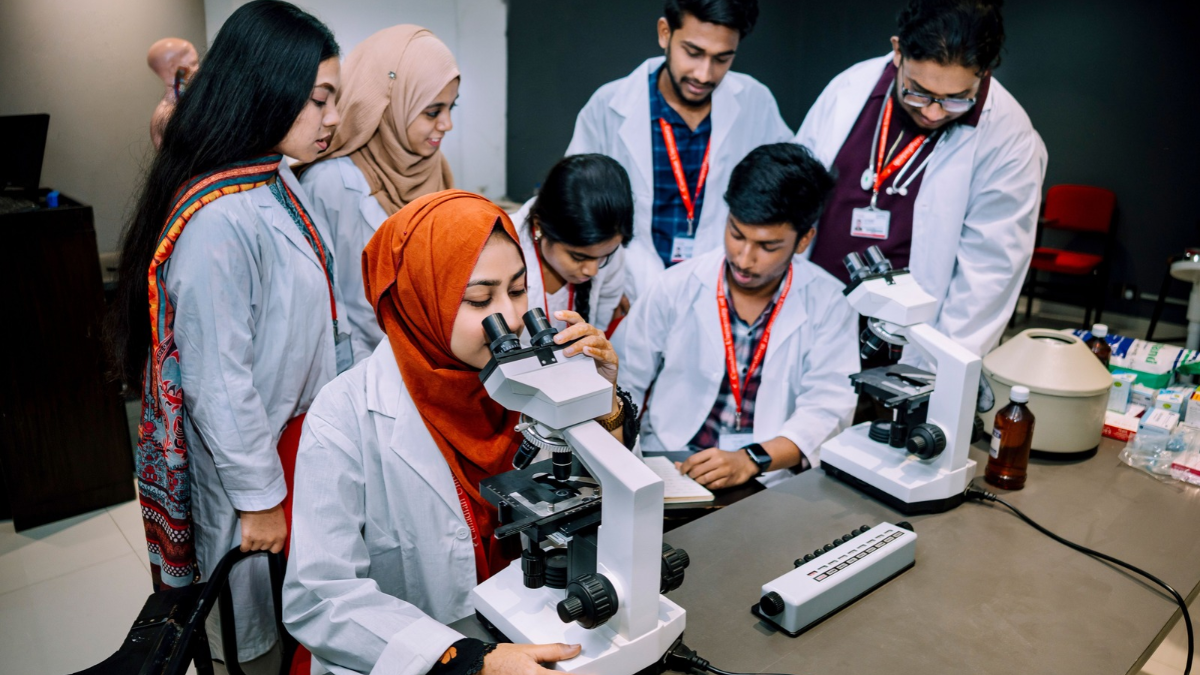
Indian medical students recognise the importance of research in strengthening healthcare, but many struggle to translate that interest into actual research projects and publications. Limited faculty mentorship, inadequate funding, insufficient research training and the pressure of academic workloads continue to restrict undergraduate research participation, according to a review published in Frontiers in Medicine on November 13, 2026.
The review, titled Understanding Research Orientation and Practice Among Undergraduate Medical/Dental Students in India: Challenges, Barriers and Enablers examines existing studies on undergraduate research engagement and evaluates national initiatives intended to develop research capabilities among medical and dental students.
The findings point to a gap between students’ willingness to pursue research and the institutional support available to help them do so.
Research interest does not translate into participation
A 2024 cross-sectional study involving 606 Indian medical students found that awareness of research opportunities did not necessarily translate into completed projects.
Although 77.23% of respondents knew about the Indian Council of Medical Research’s (ICMR) Short-Term Studentship (STS) programme, only 16.34% had applied. Just 6.4% completed their research within the prescribed period, while only 1.98% published their ICMR-related findings.
The study also examined students’ access to academic guidance. While 65.35% approached faculty members for assistance, only 46.53% found them helpful. Just 33.17% received assistance from senior students.
The findings suggest that awareness campaigns alone may not be sufficient to develop a research culture without accessible mentorship and sustained institutional support.
The 2026 review also highlights findings from individual medical colleges. At ESIC Medical College, Chennai, a study involving 241 students found that only 8.3% actively participated in research, despite 60% recognising its importance in clinical care.
At Pushpagiri Institute of Medical Sciences and Research Centre in Kerala, a study of 160 undergraduate students and interns found that 96.9% recognised the value of research, but only 12.5% had participated in ICMR-STS.
These institution-specific findings illustrate the difference between research awareness and actual engagement.
A wider institutional research gap
The challenge extends beyond undergraduate classrooms. A study examining research output across 579 Indian medical institutions, cited in a 2026 research paper, found that 57.3% had no publications.
The finding raises questions about the research environment in medical institutions and the opportunities available to students who want to participate in scientific investigation.
However, some institutions have demonstrated that structured mentorship can improve participation.
A study published in BMC Research Notes in April 2026 documented the experience of JIGNYASA, a student-led undergraduate research committee established in February 2020 at a government medical college and tertiary-care hospital in western India.
Between February 2020 and September 2025, the initiative recorded 228 student-initiated research projects, 34 grant approvals and 59 publications.

A medical student examining a report.Image credit:Pexels
The model relied on senior-student mentorship, continuous faculty guidance, research-promotion activities and regular feedback. The authors presented it as a potentially replicable approach for institutions seeking to strengthen undergraduate research without substantial additional funding.
National programmes need stronger implementation
The Frontiers review examines three initiatives: ICMR’s Short-Term Studentship programme, the National Medical Commission’s Community Health Programme and the Basic Course in Biomedical Research.
The STS programme provides undergraduate medical and dental students an opportunity to undertake short-term research under faculty supervision. The Community Health Programme introduces students to field-based health research, while the Basic Course in Biomedical Research provides structured training in research methodology.
However, the review identifies limitations, including inadequate research grants, uneven access to mentors, limited hands-on training and insufficient support for publishing findings.
It argues that medical colleges need to provide dedicated research time, accessible funding, trained faculty mentors and stronger institutional research infrastructure.
For India, developing clinician-researchers is important not only for improving medical education but also for strengthening the country’s capacity to generate evidence relevant to its own healthcare needs.
The evidence suggests that the problem is not simply a lack of interest among medical students. It is the absence of consistent opportunities and institutional structures that can turn that interest into meaningful scientific contributions.


Health
What is the Mysterious ‘Egoviruses’ in the Guts of 180 Million People
Scientists have discovered egoviruses, a previously unknown group of gut viruses potentially present in 180 million people worldwide. Their genetic connection to poxviruses could offer new clues about viral evolution.

Millions of people could be carrying a previously unknown group of disease-causing viruses in their digestive systems without ever knowing it. Scientists have now identified these microbes, named ‘egoviruses’, in human and animal gut samples, uncovering a surprising connection to one of the most important families of disease-causing viruses.
The discovery is drawing attention because egoviruses are closely related to poxviruses, the family that includes the viruses responsible for smallpox and mpox. Researchers estimate that these newly identified viruses could be present in around 180 million people worldwide. The findings offer a new perspective on the evolution of viruses and the largely unexplored microbial world inside the human digestive system.
Hidden Viral Population in the Human Gut
The discovery emerged from an extensive examination of genetic material collected from human and animal digestive systems. Rather than isolating individual viruses in laboratories, researchers used metagenomics, a technique that allows scientists to identify organisms by analysing genetic material in biological samples.
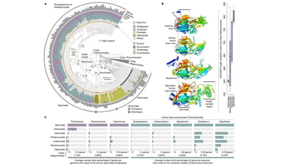
The research team, led by microbial ecologist Tom Delmont of the French National Centre for Scientific Research, identified egoviruses across samples from several countries. These included Tanzania, Nepal, Fiji, the United States and parts of Europe.
The analysis revealed six distinct groups associated with humans. One of these groups was found across multiple continents, suggesting that the viruses are not confined to a particular population or geographical region. The researchers also detected egoviruses in buffaloes, goats, chickens, deer and pigs. Their presence across different animal species points to a much wider distribution than scientists had previously recognised.
Do These Viruses Actually Infect Humans?
If egoviruses are found in human gut samples, does that mean they infect human cells? Not necessarily. The researchers suspect that these viruses may target single-celled organisms living in the digestive tract rather than human cells themselves. Genetic comparisons revealed similarities between egovirus genes and those of two groups of single-celled organisms, Parabasalia and Trichostomatia.
Some members of these groups live in animal digestive systems. Trichostomatia, for example, includes microorganisms found in the digestive tracts of ruminants, where they are involved in breaking down food. This could mean that the human gut contains viruses that interact with its microbial inhabitants rather than directly attacking human tissue. However, researchers have yet to confirm their suspected hosts through direct experiments. There is also no evidence from this study that egoviruses cause illness in humans.
Surprising Link to Smallpox and Mpox Viruses
Perhaps the most intriguing aspect of the discovery lies in the evolutionary history of egoviruses. The researchers found that these viruses are the closest known relatives of poxviruses among the groups they examined. Poxviruses are large DNA viruses that can infect humans and animals. Smallpox, eradicated in 1980, and mpox belong to this family.
The genetic relationship has led researchers to investigate whether the ancestors of poxviruses once infected single-celled organisms before evolving the ability to infect animal cells. One possibility is that an ancestral virus initially lived among microorganisms in the digestive systems of early animals. Over millions of years, evolutionary changes may have enabled some descendants to infect animal cells directly.
This remains a hypothesis, rather than a confirmed account of how poxviruses originated. Nevertheless, egoviruses offer scientists a previously unknown group of viruses through which to investigate that evolutionary transition.
What Scientists Still Need to Find Out
The discovery adds another layer to the growing understanding of the human gut, which hosts a complex community of bacteria, fungi, viruses and other microorganisms. But identifying a virus is only the beginning. Scientists still need to establish which organisms egoviruses infect, how they behave inside the gut and whether they influence the wider microbial community.
For now, the discovery of a virus group potentially carried by 180 million people raises a fascinating possibility, a large part of the human body’s viral diversity may have remained hidden simply because scientists had not yet known where to look.
Health
Six months on, Ebola outbreak in DR Congo remains uncontrolled, with over 5,500 deaths
Six months after its declaration, the Ebola outbreak in DR Congo remains uncontrolled, with over 5,500 deaths. Armed conflict, gaps in disease surveillance and limited healthcare access continue to hamper containment efforts as the Bundibugyo virus spreads across eight provinces.
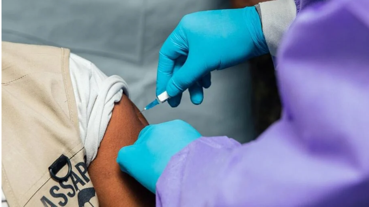
Six months after the Democratic Republic of the Congo (DRC) declared an Ebola outbreak, the disease remains out of control, with more than 11,300 confirmed cases and 5,500 deaths reported in the country. Despite an expanded international response, the outbreak continues to spread amid armed conflict, displacement and weaknesses in healthcare delivery.
The Africa Centres for Disease Control and Prevention (Africa CDC) warned on November 15 that the outbreak was still not under control. Its Director-General, Jean Kaseya, cautioned that unless transmission is interrupted, the epidemic could become the world’s worst Ebola outbreak.
According to the European Centre for Disease Prevention and Control (ECDC), the DRC had recorded 11,376 confirmed infections and 5,568 deaths as of November 15, based on data up to November 14. Eight provinces have been affected, with Ituri reporting the highest burden. The outbreak is caused by the Bundibugyo virus, a relatively rare species of Ebola virus.
Delayed detection is driving transmission
One of the biggest obstacles to containment is the inability to identify infected people and their contacts early. According to Africa CDC, more than 80% of new infections are being detected among people who were not previously listed as contacts of confirmed patients.
Contact tracing is central to Ebola control. Once an infection is confirmed, health workers identify people who may have been exposed and monitor them for symptoms during the 21-day incubation period. Failure to identify these contacts creates opportunities for further transmission.
More than 65% of deaths have occurred in communities rather than healthcare facilities, indicating that many patients are being identified too late to receive timely treatment. The World Health Organization (WHO) reported that children account for more than a quarter of confirmed infections and approximately one-third of deaths. Seven in 10 children under five who contract the disease have died.
The outbreak has expanded to 67 health zones across eight provinces, with an estimated 400–500 new cases being reported each week, according to the WHO’s November 14 update.
Conflict and insecurity disrupt response
Armed conflict and population displacement have further complicated containment. Insecurity has restricted access to affected communities, disrupted healthcare services and made surveillance and contact tracing difficult.
The WHO reported more than 80 attacks on healthcare since the outbreak began. An armed attack in Cantine-Aloya killed 66 civilians, including three health workers, while three healthcare facilities were looted. More than 1,000 contacts were subsequently lost to follow-up, and three confirmed patients could not be accounted for.
The response has also been affected by delayed payments to frontline health workers. Africa CDC has warned that inadequate resources, insufficiently trained personnel and operational difficulties are weakening efforts to contain the disease.

To help fight the spread of Ebola, UNICEF is supporting District Health Educators (DHE) in high-risk districts to conduct outreach activities with different groups, including corporate and business sectors, educational facilities, prisons, hostels and guest-houses. Photo: UNICEF/UNI790701/Bamulanzeki
Healthcare capacity expands, but gaps persist
There has been progress in strengthening the response. According to the WHO, diagnostic capacity has increased from two laboratories to more than two dozen, while nearly 2,000 treatment beds have been made available across 104 treatment and transit centres.
More than 17,000 community health workers and 46,000 community actors have received training to support surveillance, contact tracing and public awareness. Nearly 90% of registered deaths are now being managed through safe and dignified burials, reducing the risk of transmission during funerals.
However, these improvements have not translated into nationwide containment. Several initial outbreak centres, including Butembo, Mongbwalu and Nyankunde, have brought transmission under control, but new infections continue to emerge elsewhere.
The challenge of containing Bundibugyo virus
The virus itself presents another challenge. Bundibugyo is distinct from the Zaire ebolavirus responsible for the 2014–2016 West African epidemic. Vaccines developed against the Zaire species cannot be assumed to protect against Bundibugyo virus. There is currently no licensed vaccine specifically approved against this strain.
The international response therefore continues to depend on early diagnosis, isolation, supportive treatment, infection prevention and safe burial practices, alongside research into candidate vaccines and treatments.
The WHO has called for sustained, flexible funding through 2027 and improved access to communities affected by conflict. The outbreak has also prompted stronger cross-border surveillance, following infections reported in Kenya and Nigeria.
Six months into the crisis, the challenge is no longer simply mobilising an emergency response. It is ensuring that diagnostic services, trained personnel and treatment reach communities where transmission continues. Without sustained funding, improved security and effective contact tracing, the outbreak could remain a regional public health threat.
Health
Why Does the Brain’s Waste-Clearance Process Slow Down During Sleep As We Age?
A new MIT study finds that the movement of cerebrospinal fluid, which is involved in clearing waste from the brain, becomes weaker in older adults during sleep. The findings offer new insights into the relationship between ageing, sleep and brain health.

Sleep is often described as a period when the body rests and recovers. But the brain does not simply switch off when we fall asleep. It remains active, carrying out several processes that support its functioning, including the movement of fluid that helps transport waste products away from brain tissue. Now, researchers at the Massachusetts Institute of Technology (MIT) have found that this process becomes less pronounced with age. A study published in Nature Aging reports that older adults experience significantly weaker waves of cerebrospinal fluid (CSF) or brain waste clearance during sleep than younger adults.
Could changes in the brain’s waste-clearance processes help explain why the risk of cognitive decline and neurodegenerative diseases increases with age?
How Does Brain Waste Clearance Work During Sleep?
The brain produces metabolic waste as its cells carry out their normal activities. These waste products need to be transported away to maintain a healthy environment for brain cells. One of the fluids involved in this process is cerebrospinal fluid (CSF). It is a clear liquid that surrounds the brain and spinal cord, cushions them against injury, and helps maintain their chemical environment.
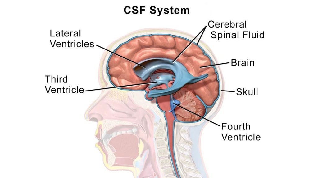
CSF also moves through spaces around brain tissue, contributing to the transport of substances and waste products. Scientists study this process in relation to the glymphatic system. The term describes a network of pathways through which fluid moves, facilitating the removal of waste from the brain.
During deep sleep, the brain produces slow, rhythmic electrical waves. These are associated with changes in blood volume, which in turn help generate waves of CSF movement. In simple terms, brain activity, blood circulation and fluid movement work together during sleep. Earlier research by MIT professor Laura Lewis and colleagues, published in Science in 2019, demonstrated how these slow brain waves are coordinated with changes in blood flow and CSF movement.
What Did the New Study Discover?
Building on their earlier work, the MIT researchers wanted to understand whether this process changes as people grow older. They recruited participants from two age groups: 18–39 years and 60–84 years. Using magnetic resonance imaging (MRI) and electroencephalography (EEG), they monitored brain activity, blood flow, and CSF movement while participants were in a scanner at night.
The researchers found that younger adults experienced stronger slow brain waves during non-rapid eye movement (NREM) sleep, accompanied by larger waves of CSF. Older participants, however, showed significantly smaller CSF waves. Importantly, the difference was not simply explained by older people sleeping for shorter periods. The researchers identified changes in the mechanisms that coordinate brain activity, blood circulation and fluid movement.
The study measured the movement and strength of CSF waves. It did not directly measure how much metabolic waste was removed from each participant’s brain. Therefore, weaker CSF waves suggest a potential impairment in a process involved in waste clearance, but do not establish that the brain actually removed less waste.
Why Does Blood Circulation Matter?
To understand what might cause the reduction in CSF movement, the researchers examined how blood vessels respond to changes in brain activity. Normally, when a part of the brain becomes more active, nearby blood vessels expand to supply additional oxygen and nutrients. This process is called neurovascular coupling.
The researchers showed participants a flickering checkerboard image to stimulate brain activity. Although older participants showed an increase in neural activity, their blood flow did not increase as much as it did in younger participants. In another experiment, participants were asked to hold their breath briefly. This raises carbon dioxide levels in the blood, normally prompting blood vessels in the brain to expand and increase blood flow.
Again, older participants showed a smaller increase in cerebral blood flow. These experiments suggest that blood vessels become less responsive with age. Since changes in blood volume help generate CSF waves during sleep, reduced vascular responsiveness may contribute to weaker fluid movement. However, the researchers found that these vascular changes did not explain the entire reduction. Other age-related factors may also be involved.
Does This Increase the Risk of Alzheimer’s Disease?
The findings are relevant to research on Alzheimer’s disease and other neurodegenerative disorders, which are associated with the accumulation of abnormal proteins and other changes in the brain. Earlier studies have linked disturbed sleep with an increased risk of cognitive decline and dementia. Researchers are interested in whether impaired fluid movement during sleep could be one of the mechanisms connecting sleep disturbances with brain health.
“We know that people age really differently. Some people age very successfully with great cognitive outcomes, and other people develop cognitive decline. If we can figure out who has great trajectories of aging and what are the factors that are associated with that, we might be able to use that to predict future outcomes,” says Laura Lewis, the Athinoula A. Martinos Associate Professor of Electrical Engineering and Computer Science, a member of MIT’s Institute for Medical Engineering and Science and the Research Laboratory of Electronics, and an associate member of the Picower Institute for Learning and Memory.
In the MIT study, some older participants showed signs of grey matter atrophy, or a reduction in brain tissue volume. This was associated with a greater decline in CSF movement. However, the study does not establish that reduced CSF movement causes grey matter loss, Alzheimer’s disease or dementia. None of the participants had been diagnosed with a neurodegenerative disorder. The researchers hope to follow some participants over time to investigate whether differences in CSF movement are associated with future cognitive outcomes.
Can the Process Be Improved?
The researchers are also exploring whether interventions could improve CSF movement during sleep. In an earlier study, Lewis’ laboratory found that short bursts of pink noise during sleep could increase CSF flow in younger adults. Pink noise is a type of sound in which lower frequencies carry more energy than higher frequencies. Whether the same intervention works in older adults has not yet been established.
Another important question is why some older participants in the new study retained CSF waves comparable to those of younger adults. Understanding what protects these individuals could help researchers identify factors associated with healthier brain ageing. For now, the findings offer a new perspective on the relationship between sleep and ageing. They suggest that the quality of sleep-related processes may matter alongside the amount of sleep a person gets. But whether improving CSF movement can actually prevent cognitive decline or reduce the risk of neurodegenerative diseases remains to be investigated.

Energy Independent by 2047? India’s Electricity Is Getting There. Its Oil and Gas Aren’t.

What is the Mysterious ‘Egoviruses’ in the Guts of 180 Million People

India’s medical students show interest in research, but mentorship and institutional support remain major hurdles

CUSAT ranks 849th globally in sustainability, tops Kerala universities

AI adoption outpaces university preparedness as Indian students turn to generative tools, FICCI-EY report finds

Indian universities lead Asia in research output per faculty, but the country’s top six all slip in QS ranking

Why Delhi’s Geography Makes It Vulnerable to Severe Air Pollution

Europe Braces for Extremes as Commission Unveils Climate Resilience Framework

NASA recruits new astronauts for future Moon Base and Mars missions

Between Lectures and Lyrics: Kabeer Is Building a Malayalam Hip-Hop Career One Self-Taught Skill at a Time
-

 Math4 months ago
The 2026 Fields Medals: Four Proofs, Four Decades-Old Problems Solved
-
EP Deep Dive: Data & Solutions4 months ago
After Kerala’s Deadliest Landslide, the Hardest Thing to Rebuild Was Childhood
-
EP Deep Dive: Data & Solutions5 months ago
West Asia Crisis: Can Kerala’s Returning Gulf Migrants Find a Future in the Green Economy?
-
Space & Physics6 months ago
India Semiconductor Mission: ‘It’s Not About Fabs. It’s About Building An Entire Ecosystem’
-
Interviews2 months ago
From Scams to Screens: How the National Stock Exchange Rewrote India’s Equity Story
-
Climate & Environment6 months ago
The Climate World Cup? How Climate Change Could Affect Player Performance at the 2026 World Cup
-
Society4 months ago
What Is Civilisational Diplomacy? Understanding India’s Newest Foreign Policy Tool
-
Society5 months ago
From Bell Labs to the Classroom: A Second Career in Teaching