
Health
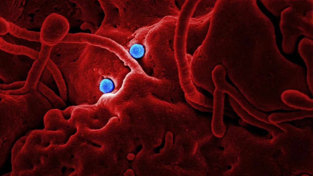
Study Reveals Essential Genes That Help Tuberculosis Survive Airborne Transmission
Tuberculosis is a respiratory disease caused by Mycobacterium tuberculosis, a bacterium that predominantly affects the lungs and spreads through droplets released by an infected person



Health
NMC Directs Medical Colleges to Set Up Patient Safety Committees
The National Medical Commission has directed medical colleges and medical institutions to establish patient safety committees, designate nodal officers and strengthen procedures to prevent avoidable harm.

Health
World Food Day: India’s Childhood Obesity Burden Raises Concerns Over Diabetes Risk
More than 41 million Indian children and adolescents aged 5–19 were living with overweight or obesity in 2025, according to the World Obesity Atlas 2026. The growing burden raises concerns about long-term health risks, including type 2 diabetes, and highlights the need for healthier food environments, better nutrition and stronger school-based interventions.

With more than 41 million Indian children and adolescents living with overweight or obesity, experts warn that unhealthy food environments could increase future risks of type 2 diabetes. New national guidance on childhood diabetes and proposed restrictions on junk food sales near schools highlight the need for preventive action.
As the world observes World Food Day on October 16, childhood nutrition is emerging as a challenge that extends beyond hunger and undernutrition. The growing prevalence of overweight and obesity among children is raising concerns about their long-term health, including their risk of developing type 2 diabetes.
India faces a particularly significant challenge. The World Obesity Atlas 2026 estimates that approximately 14.9 million children aged five to nine and 26.4 million children and adolescents aged 10–19 in India were living with overweight or obesity in 2025. Together, these figures amount to more than 41 million young people.
The report also estimates that around 1.39 million Indian children aged five to 19 had hyperglycaemia attributable to high body mass index in 2025. Hyperglycaemia refers to elevated blood glucose levels and is a health indicator associated with metabolic risk; it should not be interpreted as a count of children diagnosed with diabetes.
The figures highlight the importance of addressing childhood obesity early, before associated health risks become more difficult to manage.
Food environments and the risk of early-onset diabetes
The World Health Organization (WHO) reported in September 2026 that 400 million children and adolescents aged five to 19 worldwide were overweight in 2024, including 170 million living with obesity. The prevalence of overweight in this age group increased from 8% in 1990 to 20% in 2024.
Childhood obesity is influenced by a combination of biological, social, economic and environmental factors. Diets high in energy but low in essential nutrients, limited opportunities for physical activity and the easy availability of heavily marketed processed foods can contribute to unhealthy weight gain.
Obesity can increase insulin resistance, in which the body’s cells respond less effectively to insulin. This can raise the risk of type 2 diabetes, a condition that is increasingly important to consider in children and adolescents.
However, the relationship between food and diabetes requires an important distinction. Type 1 diabetes is an autoimmune condition in which the body’s immune system attacks insulin-producing cells in the pancreas. It is not caused by eating sugary foods or being overweight. Type 2 diabetes develops through different mechanisms and is associated with factors including excess body weight, genetics and physical inactivity.
The two conditions require different approaches to prevention and treatment.
India introduces a national childhood diabetes framework
In a significant policy development, the Union Ministry of Health and Family Welfare released a comprehensive guidance document on diabetes mellitus in children on May 3, 2026.

Diabetic checkup.Representational image.Image credit:Pexels
The framework outlines a structured approach to screening, diagnosis, treatment and long-term management, linking community-level identification with district hospitals and medical colleges. It also provides for free-of-cost care at public health facilities, including insulin therapy, blood glucose monitoring supplies and follow-up services.
The guidance promotes awareness of the early warning signs of type 1 diabetes through the “4Ts”: Toilet, Thirsty, Tired and Thinner. These refer to frequent urination, excessive thirst, unusual tiredness and unexplained weight loss.
The initiative is important because timely diagnosis and access to insulin can be lifesaving for children with type 1 diabetes. It also highlights the need for health systems to identify and manage diabetes before complications develop.
Schools become a focus of food policy
India is also considering measures to improve the food environments surrounding schools. A Reuters report published on September 24, 2026, said the country was moving towards restrictions on the sale of foods high in fat, salt and sugar within a 50-metre radius of schools.
The proposed measures would affect vendors and shops selling products such as sugary drinks and packaged snacks near educational institutions. The proposal reflects growing concern about the availability of unhealthy foods in places where children spend much of their day.
If implemented effectively, school-focused measures could form part of a wider strategy that includes healthier school meals, accessible drinking water, nutrition education, physical activity and clearer food labelling.
However, restrictions alone cannot address the problem. Families also need affordable access to fruits, vegetables, pulses and other nutritious foods. Public-health measures must account for household budgets and the realities of food access in different communities.
Beyond calories
World Food Day 2026, themed “Innovate today. Nourish tomorrow,” offers an opportunity to reconsider what food security means for children. Access to sufficient calories is essential, but the nutritional quality of food also matters.
Reducing the future burden of type 2 diabetes requires action across healthcare, education, food policy and community environments. At the same time, children living with type 1 diabetes need timely diagnosis, uninterrupted insulin supplies and long-term medical support
Health
Why Some People Age Better Than Others May Be Written in Their Blood
A study links patterns in blood proteins and immune cells to long-term disease risk, offering new insights into why some people age more healthily than others.

Ageing does not affect everyone in the same way. While some people remain relatively healthy into their later years, others develop several chronic illnesses despite being of a similar age. Scientists are investigating whether part of this difference can be traced to the way the immune system changes over a lifetime.
A study published in Immunity, offers a new clue. Researchers found that the balance between two populations of immune cells was associated with people’s long-term risk of disease and death. They also developed a way to estimate this balance using proteins circulating in the blood.
The findings could help scientists identify biological changes associated with unhealthy ageing before symptoms appear. Why do some people’s immune systems remain resilient while others develop patterns associated with declining health?
Ageing Leaves Marks on the Immune System
The immune system changes as people grow older. Its ability to respond to unfamiliar threats can weaken, while some immune cells become more prominent after years of exposure to infections and other stimuli. These changes can influence how the body responds to disease.
To investigate these differences, researchers examined approximately 12.4 million immune cells collected from 2,609 adults aged 20 to over 90 across eight study groups. Their analysis focused on two populations of T cells, which are white blood cells involved in identifying and responding to threats. The cells were distinguished by their production of granzyme K and granzyme B, proteins that help coordinate immune responses and destroy infected or abnormal cells.
The researchers found that older adults did not share a single pattern of immune ageing. Instead, their immune systems varied considerably, with some showing a greater presence of cells associated with granzyme B and others displaying a different balance.
That variation mattered when the team examined participants’ health outcomes. An immune profile dominated by granzyme B-producing cells was associated with a higher risk of several chronic conditions and death over the following years. The finding suggests that a person’s immune system may reveal aspects of their health trajectory that their age alone cannot capture.
What Blood Proteins Can Reveal
Counting specific immune cells requires specialised laboratory techniques. The researchers therefore investigated whether the proteins circulating in blood could offer an alternative way to estimate the same underlying pattern. They used a smaller dataset linking immune-cell measurements with blood-protein levels to develop a computer model. This model was then applied to data from approximately 50,000 participants in the UK Biobank, a long-running research resource that tracks health and biological information.
The team compared the predicted immune profiles with participants’ subsequent health records. Those whose profiles indicated a greater abundance of granzyme B-producing cells had higher risks of death and conditions including type 2 diabetes, hypertension, liver disease and kidney disease. The researchers followed health outcomes over an extended period, allowing them to investigate whether the blood-protein patterns were associated with future illness rather than simply reflecting differences observed at one point in time.
Could This Lead to an Early-warning Test?
One potential application is a blood test that helps identify people whose immune systems show signs associated with poorer long-term health. Many chronic diseases develop over years. Detecting biological changes before a person experiences noticeable symptoms could eventually give researchers and clinicians an additional way to assess risk and investigate preventive strategies.
The research team is working towards a simpler testing approach that could use standard laboratory equipment. At present, however, the findings represent a research advance rather than an established clinical test for healthy ageing. A useful test would need to do more than identify a statistical association. It would have to demonstrate that its predictions are sufficiently accurate for individual patients, work reliably across different populations and provide information that improves clinical decisions.
An Association That Needs Explanation
A greater abundance of granzyme B-producing cells could reflect underlying immune stress or other biological processes associated with disease. Researchers will need further investigation to establish whether these cells contribute directly to the development of chronic conditions or primarily act as indicators of changes occurring elsewhere in the body.
The results also require validation before the proposed protein signature can be used widely. A model developed from one set of participants may not perform equally well in people with different genetic backgrounds, lifestyles, environmental exposures or existing health conditions.
For ageing research, the larger opportunity lies in understanding why people follow different biological trajectories. A reliable marker could help scientists study these differences and identify when intervention might be most effective. The immediate achievement is a potential way to read some of those differences through blood. Whether that information can translate into longer, healthier lives will depend on the evidence that follows.


NMC Directs Medical Colleges to Set Up Patient Safety Committees

The Monsoon’s New Rhythm: Less Rain, More Intense Downpours

World Food Day: India’s Childhood Obesity Burden Raises Concerns Over Diabetes Risk

COP17: Can the World Still Meet Its Biodiversity Goals by 2030?

Chandigarh’s Sukhna Wetland Gets Ramsar Recognition, Taking India’s Count to 103

From Scams to Screens: How the National Stock Exchange Rewrote India’s Equity Story

Knowing When to Think for Ourselves: AI, Intuition and the Education of Discernment

After a Scorching Summer, Delhi’s Children Are Slow to Return Outdoors

Two Chemists Win Nobel Prize for Solving a Century-Old Puzzle: Why Life Picks One Hand Over the Other

India’s AI Moment Rests on More Than Code

-

 Math3 months ago
The 2026 Fields Medals: Four Proofs, Four Decades-Old Problems Solved
-
EP Deep Dive: Data & Solutions3 months ago
After Kerala’s Deadliest Landslide, the Hardest Thing to Rebuild Was Childhood
-
EP Deep Dive: Data & Solutions3 months ago
West Asia Crisis: Can Kerala’s Returning Gulf Migrants Find a Future in the Green Economy?
-
Space & Physics5 months ago
India Semiconductor Mission: ‘It’s Not About Fabs. It’s About Building An Entire Ecosystem’
-
Interviews4 weeks ago
From Scams to Screens: How the National Stock Exchange Rewrote India’s Equity Story
-
Climate5 months ago
The Climate World Cup? How Climate Change Could Affect Player Performance at the 2026 World Cup
-
Society3 months ago
What Is Civilisational Diplomacy? Understanding India’s Newest Foreign Policy Tool
-
Society4 months ago
From Bell Labs to the Classroom: A Second Career in Teaching